Whoops!

This website no longer supports this web browser. Use one of these browsers to get the best possible experience.

All-Star Surgical Precision Defeats Brain Tumors

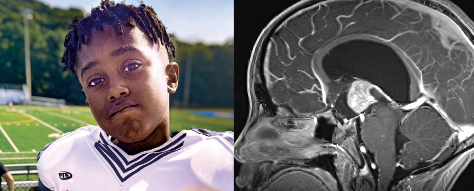
It started innocently enough: nine-year-old Kevin was having regular headaches. They were cramping his style on the basketball court and the football field. When the headaches grew more frequent over the course of a month, his mother, Jhanell, took him to the pediatrician and then to the eye doctor, since the headaches were centered behind his eyes. The eye doctor saw there was swelling of the optic nerves, so she sent him to nearby hospital. There they found a mass in his head, and Kevin was sent to the specialists at Connecticut Children’s, where a series of scans discovered two tumors in his brain.

“When they told me that,” Jhanell says, “I almost had a nervous breakdown, but then I looked at Kevin and he was looking at me and his dad for our reactions. I had to keep it together a little bit because he was there needing me to be strong.”

The good news was the tumors were not cancerous. They were the result of a genetic condition called tuberous sclerosis, which causes the body to generate multiple tumors throughout the body. Several types of lesions can form in the brain, and they can sometimes grow large enough to affect brain function.

Kevin's surgery was entirely successful.

In Kevin’s case, the tumors in his brain had formed in the ventricles, the chambers in the center of the brain that produce cerebrospinal fluid. One of the tumors was blocking the outlet of the ventricles, so the continuously produced fluid was backing up, causing the chambers upstream to enlarge. That, in turn, was putting pressure on his brain, causing the headaches and eye problems.

To address that problem, neurosurgeon David Hersh, MD, had to insert a tube through a small hole in the skull and draw out the excess fluid to release pressure on the brain. Then he used a clear cylindrical retractor to gently create a path to the tumor deep within the brain. This allowed him to use long, delicate microinstruments to remove the tumor.

The first tool that Dr. Hersh used was microbipolar forceps, which precisely and accurately apply heat to coagulate blood vessels that feed the tumor. Next, he used microscissors, with the pivot very near the tip and blades that are slightly larger than a grain of rice, to cut the coagulated vessels. Once this was complete, he was able to use a microdissector to “disconnect” the tumor from the surrounding healthy tissue. Finally, once the tumor was free, he used special tumor microforceps to pull the tumor out. These have tiny toothed discs on the tips to help grip the tumor.

The second brain tumor and the others in his body can be controlled with a medication that inhibits their growth.

This procedure demonstrates why it’s so important to have the right tools for the job. The finer and more precise the tool, the less likely any collateral damage. Tools with specialized tips give surgeons the ability to deal with a wide range of situations. And tools of varying lengths mean they can reach the right spot with maximum control.

Happily, Kevin’s surgery was entirely successful, and the medication is keeping his other tumors from growing, so he is getting back to being his old self. Football will have to wait a few months, but once the healing is complete, Kevin will be on the field again.

Aesculap Micro Tools

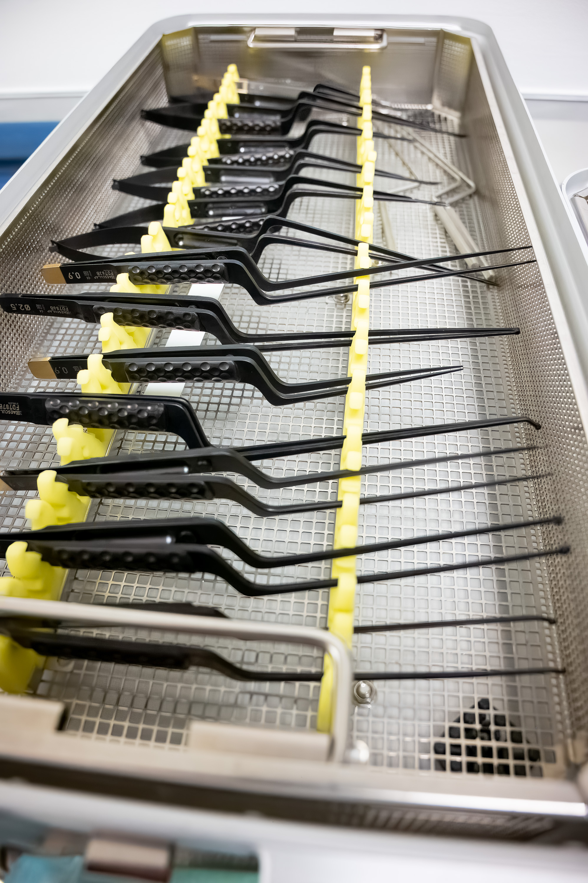
Connecticut Children’s recently acquired a full set of Aesculap micro tools for neurosurgery, thanks to the Walden W. and Jean Young Shaw Foundation. These tools come in a variety of shapes and sizes, each for a very specialized task or situation. Various lengths means having the exact right tool, minimizing the risk of damaging brain tissue. One of the tools in the set is a pair of micro tumor forceps, which feature many of the benefits common to the whole set.

Secure Grip

The “golf ball” texture on the handles of the forceps ensure the surgeon can get a secure grip on the tool—crucial when working on a child’s brain.

Improved Visibility

The bayonet shape of the tool, with the handle lower than the tip, means that the surgeon’s hand is not blocking his or her view of the working end of the tool.

Wear & Glare Resistant

The tools have a diamond dust coating that has two advantages: it makes the surface harder and more wear resistant, and the black coating on the tool ensures that bright surgical lights aren’t reflected into the surgeon’s eyes, as they could be with a brighter surface.

Improved Removal

The micro tumor forceps are designed to grab and remove tumor tissue. To that end, they have serrated rings on the tip, which provide a firm grip on the tissue.

Latest Articles

Christian

Revolutionizing Respiratory Care: New Advances Are Transforming Lives for PCD Patients

Learn More
Long-time donors Debra and Louis Chênevert and their daughters, Sophie Chênevert-Schilke and Lisa Chênevert-Krause.

$3.5 Million Gift Establishes the Chênevert Family Advanced Cellular and Gene Therapy Unit

Learn More
A toddler is seen by Audiology. She holds a distraction toy, provided by Child Life.

When Hearing Hurts: Transforming Lives Through Sound

Learn More
All Articles